THE BACK MUSCLES ANATOMY
ERECTOR SPINAE GROUP
INTERMEDIATE LAYER
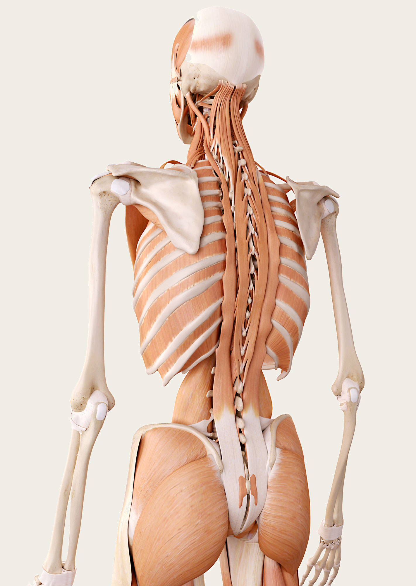 The intermediate layer of the intrinsic back muscles lies deep to the splenius group of muscles but superficial to the other intrinsic back muscles. It is formed mainly from the erector spinae group, which is the main extensor of the vertebral column.
The erector spinae muscles, also known as sacrospinalis and extensor spinae, run more or less vertically and support the entire spinal column. The group is attached from the sacrum to all five lumbar vertebrae as well as to the bottom of the two thoracic vertebrae.
At the thoracic spine, the muscular fibers split into the: iliocostalis, longissiumus and spinalis. In the cervical region it is covered by the nuchal ligament and in the thoracic and lumber region by the thoracolumbar fascia.
The muscles of the erector spinae group are the strongest muscles in the back, and they take on most of the work to support the spine. The deep back muscles are both extensors and rotators of the axial skeleton.
The erector spinae muscle group is divided into three distinct columns or fascicles, from medial to lateral:
- Spinalis muscles
- Longissimus muscles
- Iliocostalis muscles
Each fascicle can be further subdivided according to the region of the vertebral column that it inserts into. For example, we refer to the lumborum, thoracis, cervicis, and capitis for those that insert respectively in the lumbar, thoracic and cervical vertebrae or the base of the skull.
The muscles are innervated by the lateral branches of the dorsal rami arising from the cervical, thoracic, and lumbar spinal nerves.
The main function of the erector spinae group is to move the vertebral column:
- Bilateral contraction of these muscles extends the spine.
- Unilateral contraction causes lateral flexion (ipsilateral).
- They also help to maintain posture by stabilizing the spine on the pelvis during walking.
TRANSVERSOSPINALES GROUP
DEEP LAYER
The third layer of the intrinsic back muscles is formed from the transversospinales group, located underneath the erector spinae. These short, oblique muscles run obliquely and medially from the transverse process of the vertebra below to the spinous process, filling the groove of the spinous process.
There are three subdivisions of this muscle group based on their layers.
From superficial to deep, these include:
- Semispinalis
- Multifidus
- Rotatores
Similar to the previous muscle groups, the transversospinales can be further subdivided according to the region of the vertebral column that they insert into: the capitis, cervicis, thoracis, and lumborum.
DEEPEST LAYER
The deepest layer of the vertebral column is formed by the minor deep intrinsic muscles of the:
INTERSPINALES
It spans between adjacent spinous processes. They are divided into interspinales cervicis, interspinales thoracis and interspinales lumborum.
The function of the interspinales muscles is to stabilize the vertebral column and assist in the extension of the cervical and lumbar spines.
INTERTRANSVERSARII
As the name suggests, the intertransversarii muscles are small fascicles that expand between the transverse processes of adjacent vertebrae. These muscles are present at all levels of the vertebral column, but they are most developed in the cervical region. They assist with lateral flexion and with stabilizing the spinal column.
The anterior and posterior rami of the spinal nerves emerge along the length of the muscle to initiate motor activity. As a result, the intertransversarii muscles stabilize the spine (bilateral contraction) and the lateral flexion of the spine (unilateral contraction).
LEVATORES COSTARUM
The levatores costarum muscles are minor deep back muscles that are not well-developed. When present, they originate from the transverse processes of C7–T11, and attach to the rib immediately below.
They are innervated by the posterior rami of the spinal nerves arising along the thoracic vertebrae.
They act to elevate the ribs and rotate the thoracic spine.
INTERMEDIATE LAYER (Extrinsic Muscles)
Muscles: serratus posterior superior and serratus posterior inferior muscles.
Innervation: Intercostal nerves.
Functions: Movement and stabilization of the vertebral column and thorax. Aiding respiration.
The muscles of the back can be divided into three groups – superficial, intermediate and deep or intrinsic. The intermediate group is associated with movements of the thoracic cage and contains two muscles: the serratus posterior superior and serratus posterior inferior.
These muscles run from the vertebral column to the rib cage, and assist with elevating and depressing the ribs. They are thought to have a slight respiratory function.
Note: See The Shoulder Girdle course, part of the Functional Anatomy Certification for details on the serratus posterior superior and serratus posterior inferior muscles.
THE SUPERFICIAL LAYER (Extrinsic Muscles)
The muscles of the back can be divided into three groups: superficial, intermediate and deep or intrinsic. The superficial back muscles are classified as extrinsic muscles; they are situated underneath the skin and superficial fasciae. They originate from the vertebral column and attach to the bones of the shoulder girdle as the clavicle, scapula and humerus.
The trapezius and the latissimus dorsi are the most superficial muscles that form this group, with the trapezius covering the rhomboids and levator scapulae.
They are mainly associated with movements of the shoulder.
Muscles
The muscles that form this superficial group are:
- Latissimus dorsi
- Trapezius*
- Rhomboid (major and minor)*
- Levator scapulae*
Innervation
Ventral rami of the cervical nerves (except the trapezius–accessory nerve).
Functions
Moving the scapula in several directions and holding it in place.
* For more details on the trapezius muscle, see The Shoulder Girdle course, part of the Functional Anatomy Certification.
